冰冻三尺 非一日之寒
积土成山 非斯须之作
我是帅磊,本硕均在河海大学,目前河海大学信息工程学院电子信息专业研三在读。
我的主要工作是在蒋俊锋教授的指导下进行医学图像处理。我参与了常州市图形与骨科植入物数字技术重点实验室的多个项目和任务。我的工作涉及目标检测、图像分割、关键点识别和2D/3D配准。
我的GitHub主页👉GitHub·ThreeStones 与 CSDN主页👉SL1029。
最新
- 2024.10.14 论文《ABLSpineLevelCheck: Localization of Vertebral Levels on Fluoroscopy via Semi-supervised Abductive Learning》被BIBM2024接收为short paper(接收率:21%)。
主要项目
- 2024.12-2025.01 基于椎节细节分割与数学优化的椎弓根螺钉自动规划
- 2024.11-2024.12 基于改进yolov5的术中X线片旋转检测+标志点识别
- 2024.08-2024.09 基于ffmpeg的多线程音视频解码编码系统
- 2024.04-2024.07 基于对比学习的术中X线片骨折识别
- 2023.12-2024.04 基于溯因学习的术中X线片椎骨识别(BIBM2024)
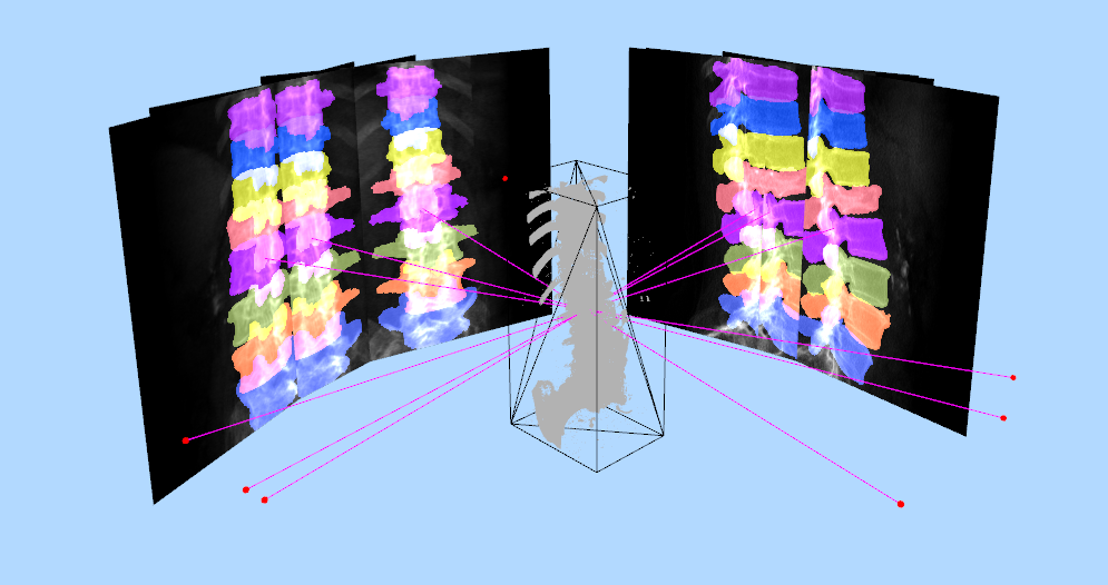
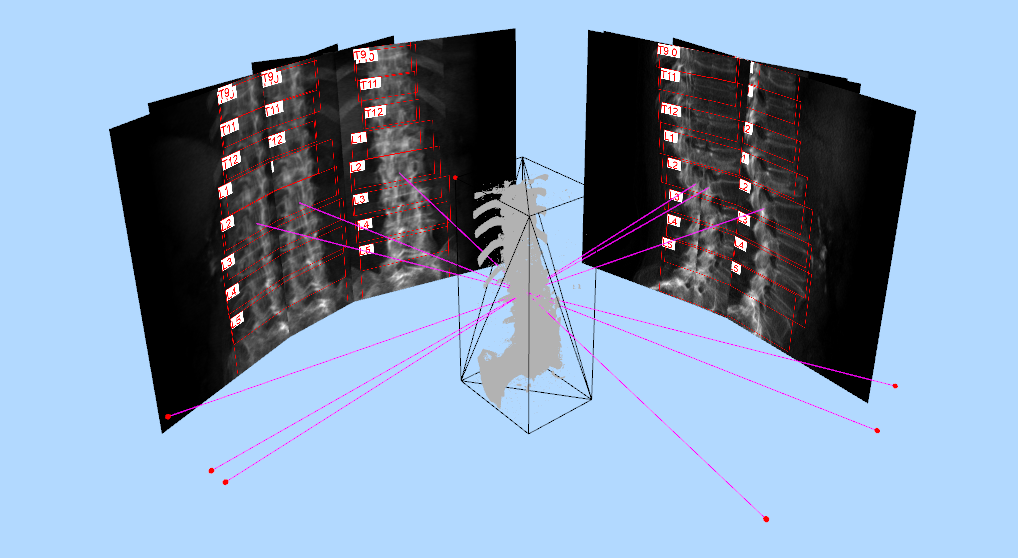
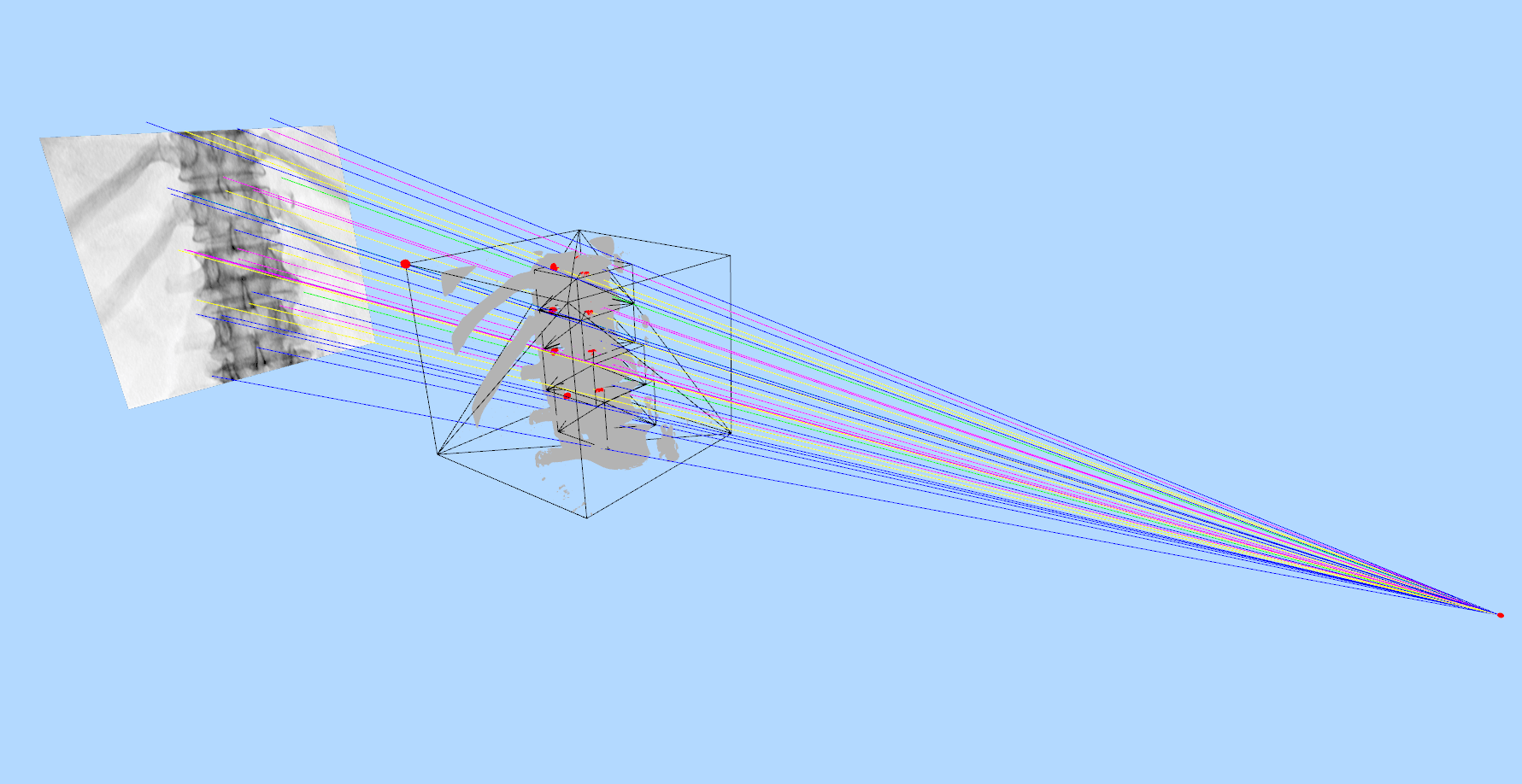
- 2024.01 脊柱DRR生成与标注 python代码 和 C++代码
- 2023.11-2023.12 基于nnunet的椎骨分割
- 2023.07-2023.08 基于HRNet的椎骨关键点检测
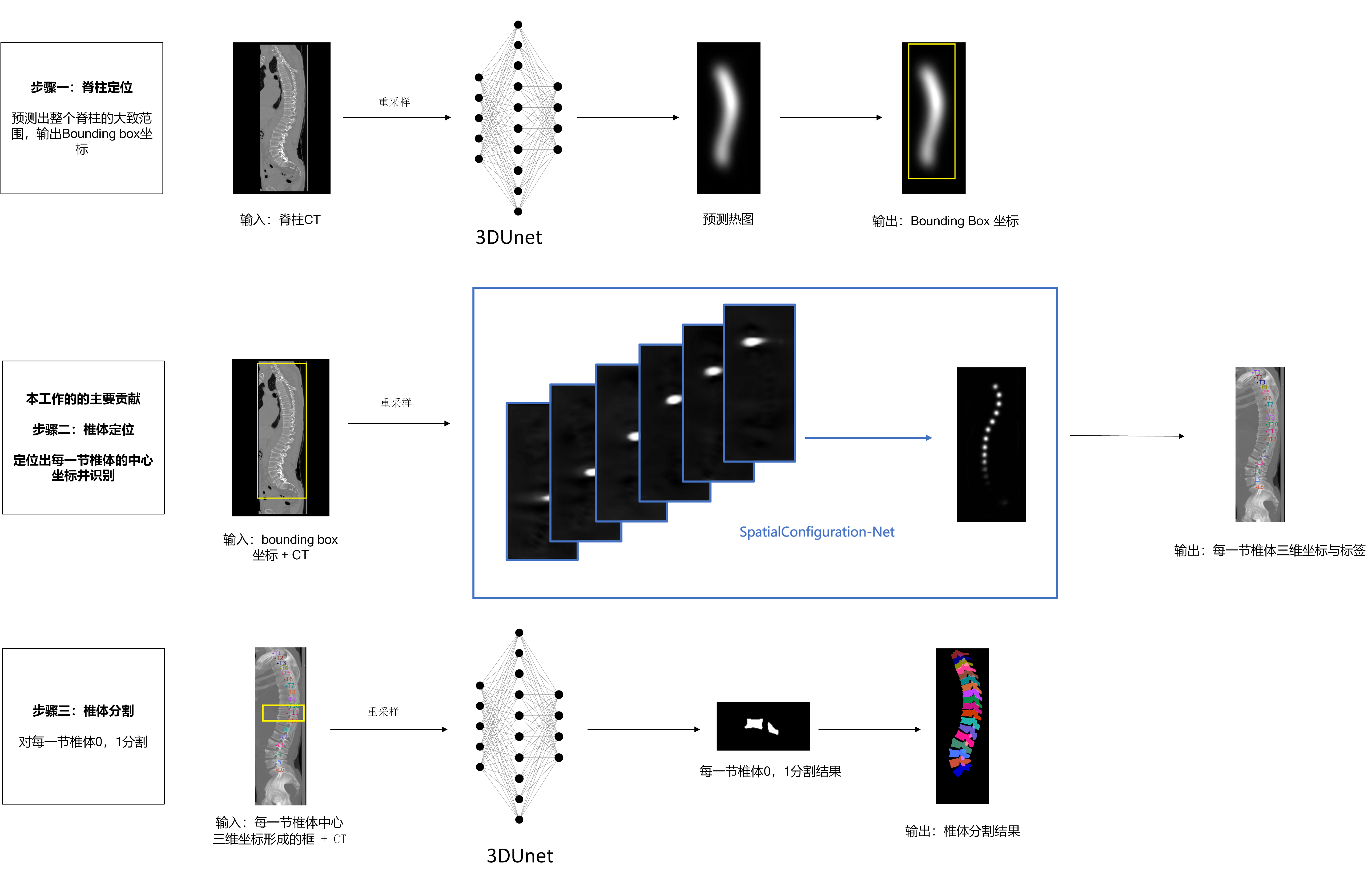
- 2023.02-2023.07 脊柱CT椎骨分割与定位
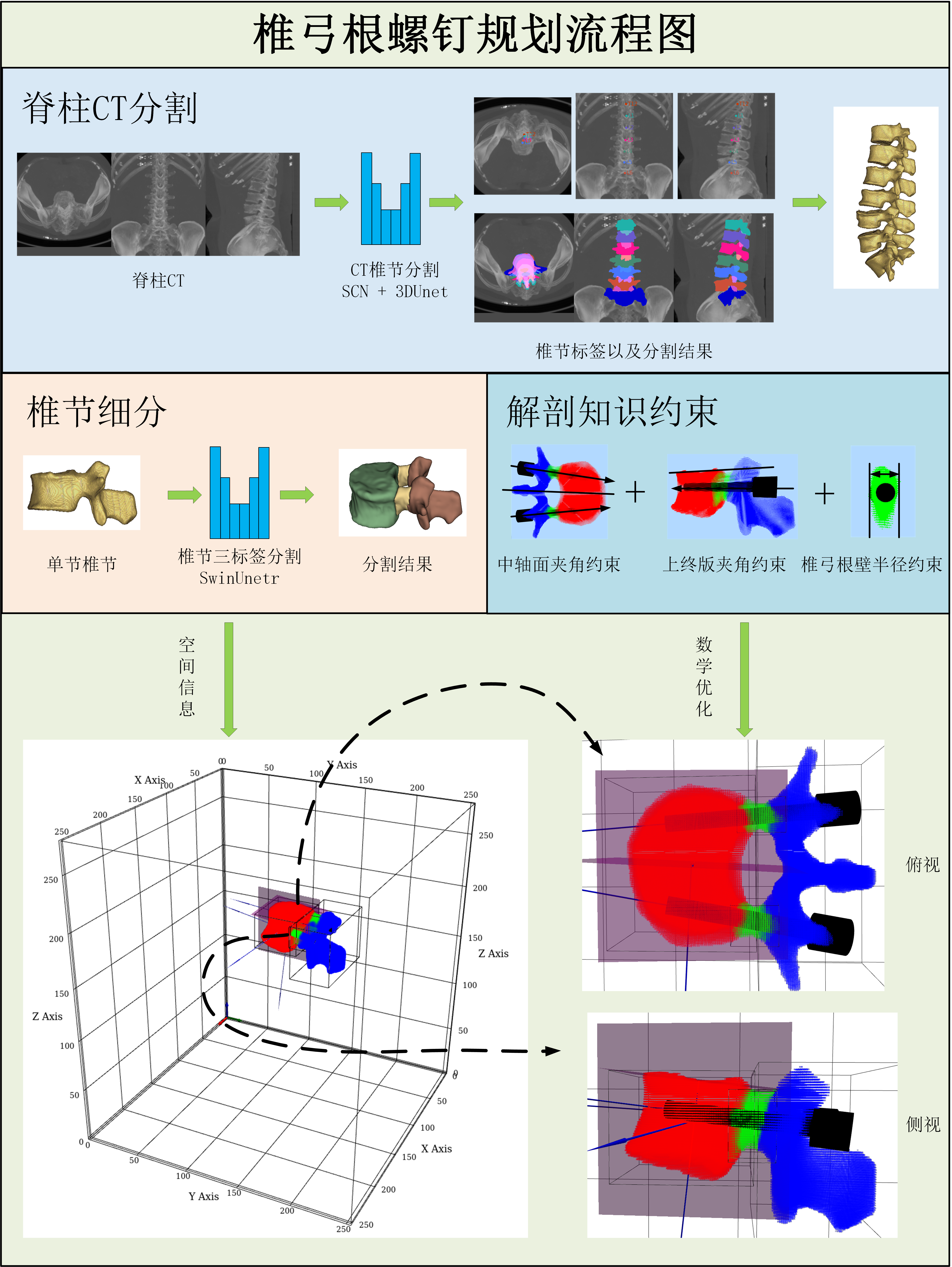
在前面CT分割项目的基础上,对每一节椎节进行三标签的分割,将椎节分割为椎体、椎弓根、其他三部分。在此基础上进行螺钉规划,具体主要根据数值优化的方法,来约束螺钉的起点、方向、大小和长度。 约束条件主要来自于临床医生手术总结的经验,例如螺钉和上终板的夹角、螺钉和中轴面的夹角等。
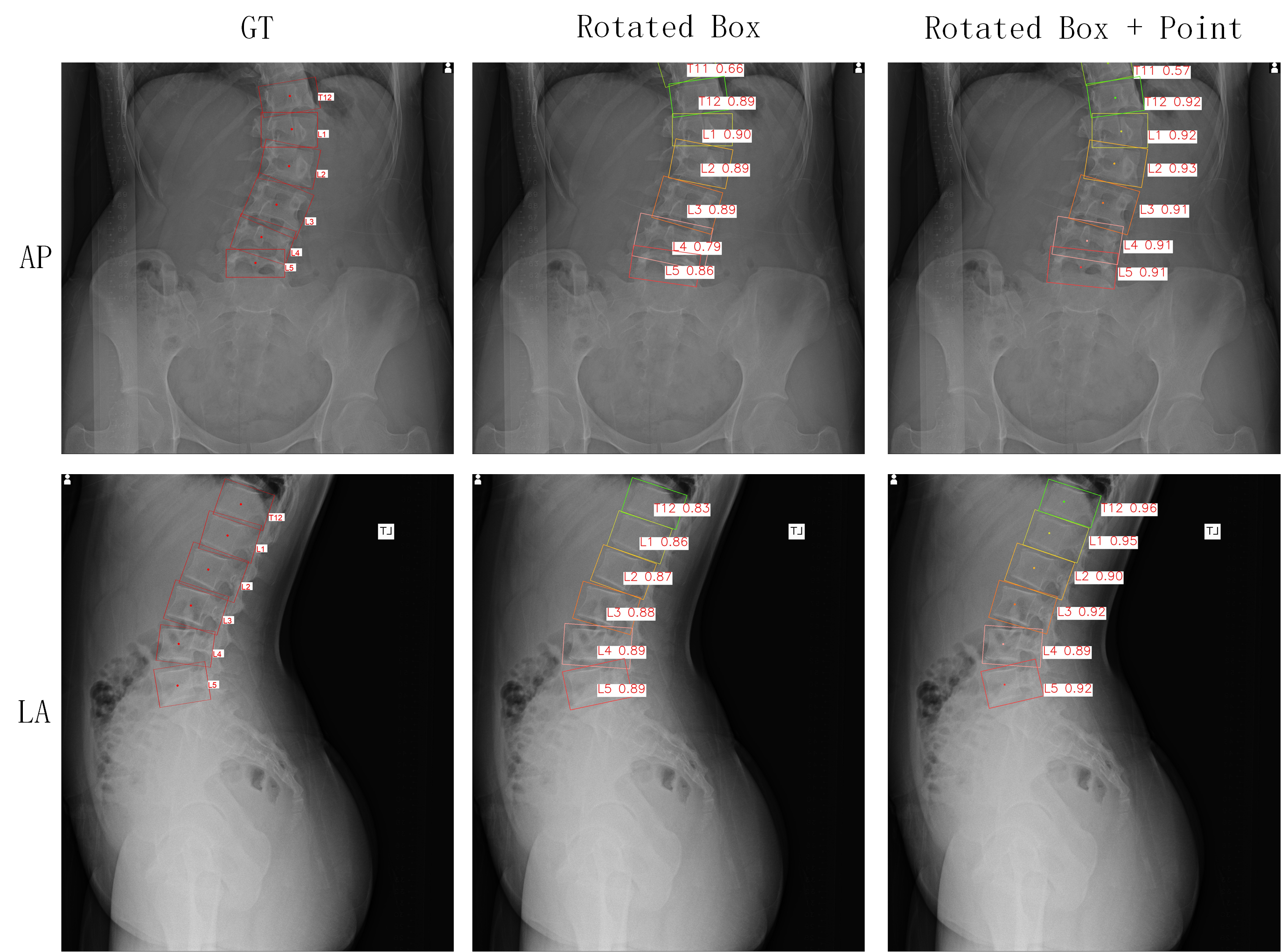
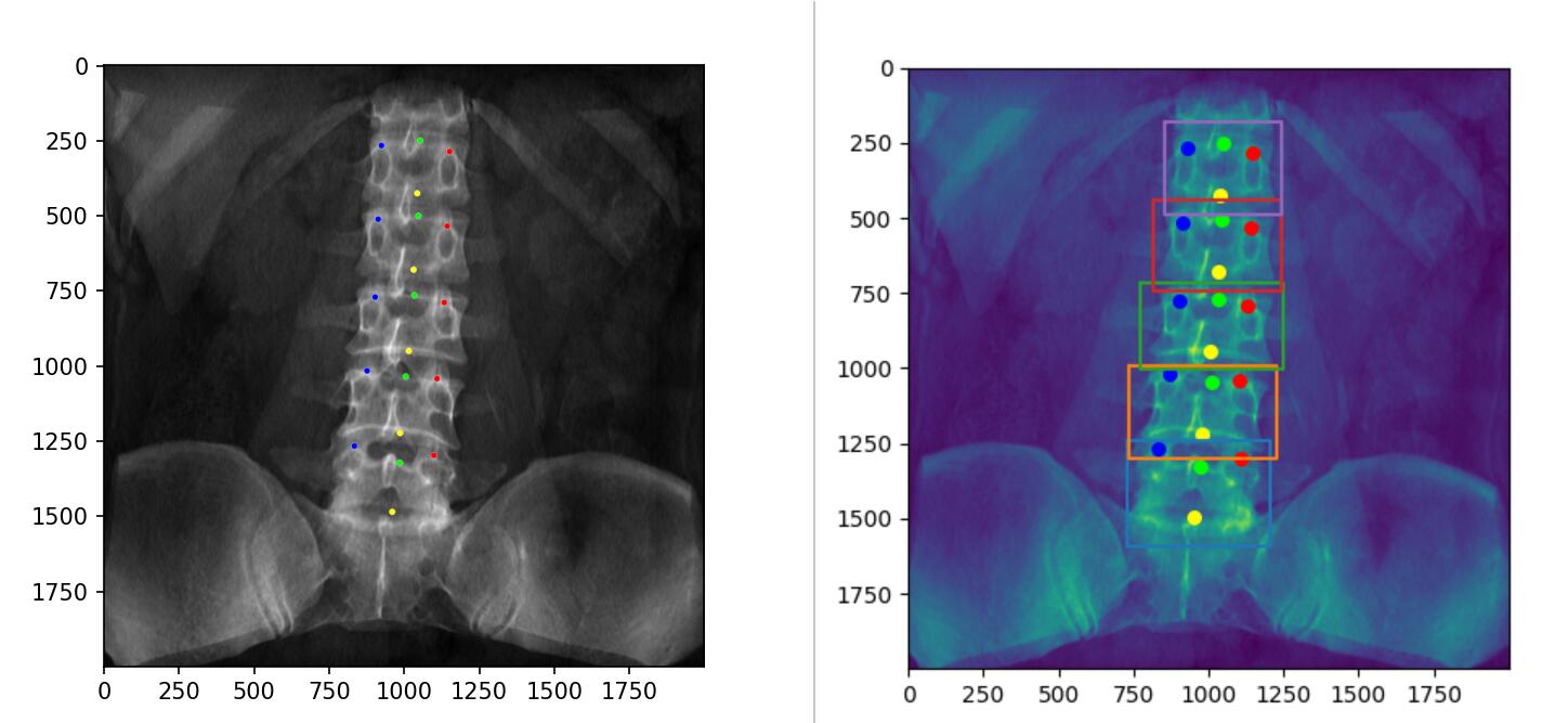
改进术中X线片检测,将水平检测框改进为旋转检测框,同时为了提升2D-3D配准准确率,加入椎骨中心点识别,改进yolov5同时输出旋转检测框和关键点。

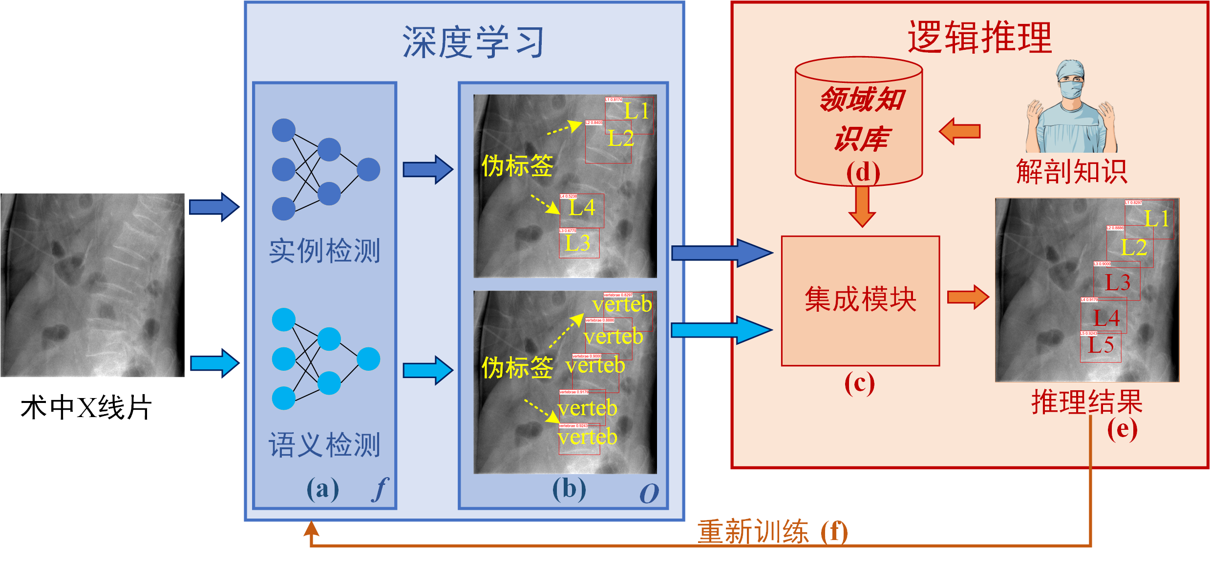
深度学习在x射线图像中的椎骨定位方面显示出了良好的效果,尽管它在成分泛化、数据效率和可解释性方面存在不足。为了解决这个问题,我们引入了一种溯因学习机制,属于神经符号范式。 最初,未注释的脊柱透视图像由神经网络推理,以推断椎体定位的伪标签。随后,这些伪标签通过由一阶逻辑子句组成的知识库进行溯因推理。然后利用推理的结果对网络进行再训练。 此外,我们提出了一种集成技术,将椎体语义检测与实例检测相结合。为了进一步提高性能,我们合成了一个数据集,并对BUU数据集进行了注释,用于网络预训练。消融研究证实了我们的方法中提出的组件的有效性。 此外,对比分析表明,我们的方法显着超越了领先的目标检测算法,以最少的注释表现出卓越的性能。

)T8%5B%5B4BDC1GRR%7DZ@AQ$%60CK.png)
Frozen three feet is not a day's cold
Making mountains from mounds is not done in a short time
Hi, I am Shuai Lei,Both my master's and master's degrees are in Hohai University. At present, I am a research junior student in the School of Information Engineering, Hohai University.
I primarily worked under the guidance of Prof. JunFeng Jiang on medical image processing. I participated in several projects and tasks at the ChangZhou Key Laboratory of Digital Technology on Graphics and Orthopaedic implants. My work involved object detection、image segmentation、keypoints recognition and 2D/3D registration.
My github home page👉GitHub·ThreeStones and CSDN home page👉SL1029。
News
- 2024.10.14 The paper《ABLSpineLevelCheck: Localization of Vertebral Levels on Fluoroscopy via Semi-supervised Abductive Learning》was accepted by BIBM2024 as a short paper(Acceptance rate:21%).
Main Projects
- 2024.12-2025.01 Automatic pedicle screw planning based on detailed segmentation of vertebral and mathematical optimization
- 2024.11-2024.12 Intraoperative X-ray rotation detection + landmark point recognition based on improved yolov5
- 2024.08-2024.09 Multithreaded audio and video decoding coding system based on ffmpeg
- 2024.04-2024.07 Contrastive Learning for Fracture recognition in Intraoperative Radiographs
- 2023.12-2024.04 Vertebrae Recognition on Intraoperative Radiographs Using Abductive Learning(BIBM2024)
- 2024.01 Spine DRR generation and annotation python code 和 C++ code
- 2023.11-2023.12 Vertebra segmentation based on nnunet
- 2023.07-2023.08 Keypoint detection of vertebrae based on HRNet
- 2023.02-2023.07 Vertebral segmentation and localization in spine CT
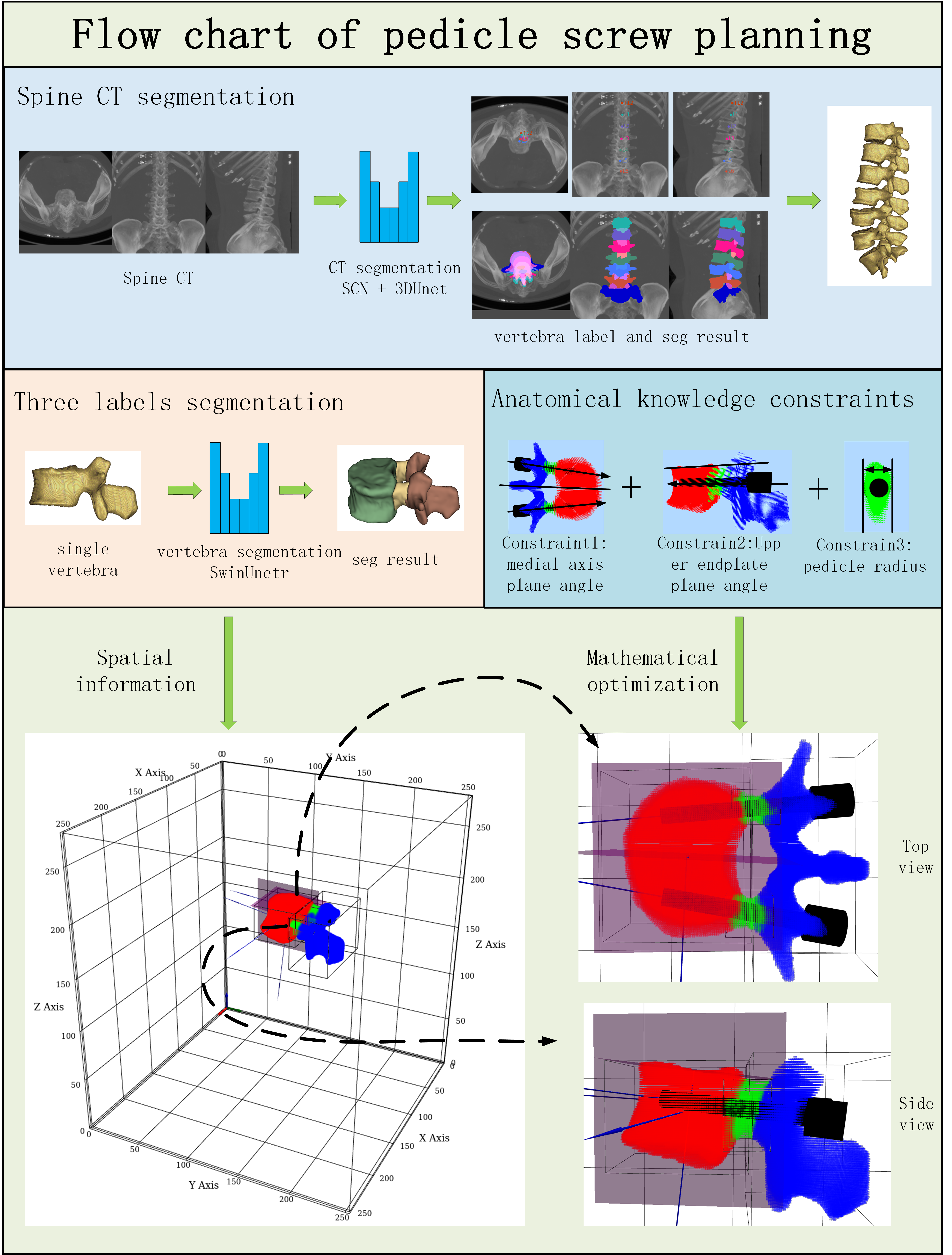
On the basis of CT segmentation items, each vertebral segment was segmented with three labels. The vertebral segment is divided into the vertebral body, pedicle and other three parts. On this basis, screw planning is carried out, which mainly restricts the starting point, direction, size and length of screws according to the numerical optimization method. The constraints mainly come from the experience of the clinician, such as the Angle between the screw and the upper end plate, and the Angle between the screw and the axial surface.

The intraoperative X-ray detection was improved, the horizontal detection frame was changed to the rotating detection frame, and the recognition of vertebral center point was added to improve the accuracy of 2D-3D registration. Improved yolov5 to output both the rotation check box and key points.

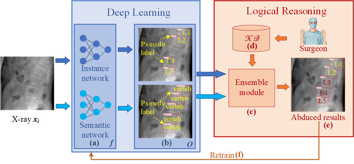
Deep learning has demonstrated promising efficacy in the localization of vertebrae within X-ray imagery, although it is recognized for its deficiencies in compositional generalization, data efficiency, and interpretability. To address this issue, we introduce an abductive learning mechanism, situated within the neuro-symbolic paradigm, tailored for semi-supervised vertebral localization. Initially, unannotated spinal fluoroscopic images are processed by the networks to infer pseudo-labels for vertebra localization. Subsequently, these pseudo-labels undergo abductive reasoning via a knowledge base comprised of first-order logical clauses. The networks are then retrained utilizing the abducted outcomes. Additionally, we propose an ensemble technique that amalgamates semantic detection of vertebral levels with instance detection. To further augment performance, we have synthesized a dataset and annotated the BUU dataset for network pretraining. Ablation studies validate the efficacy of the proposed components in our methodology. Furthermore, comparative analyses reveal that our approach significantly surpasses leading object detection algorithms, exhibiting superior performance with minimal annotations.

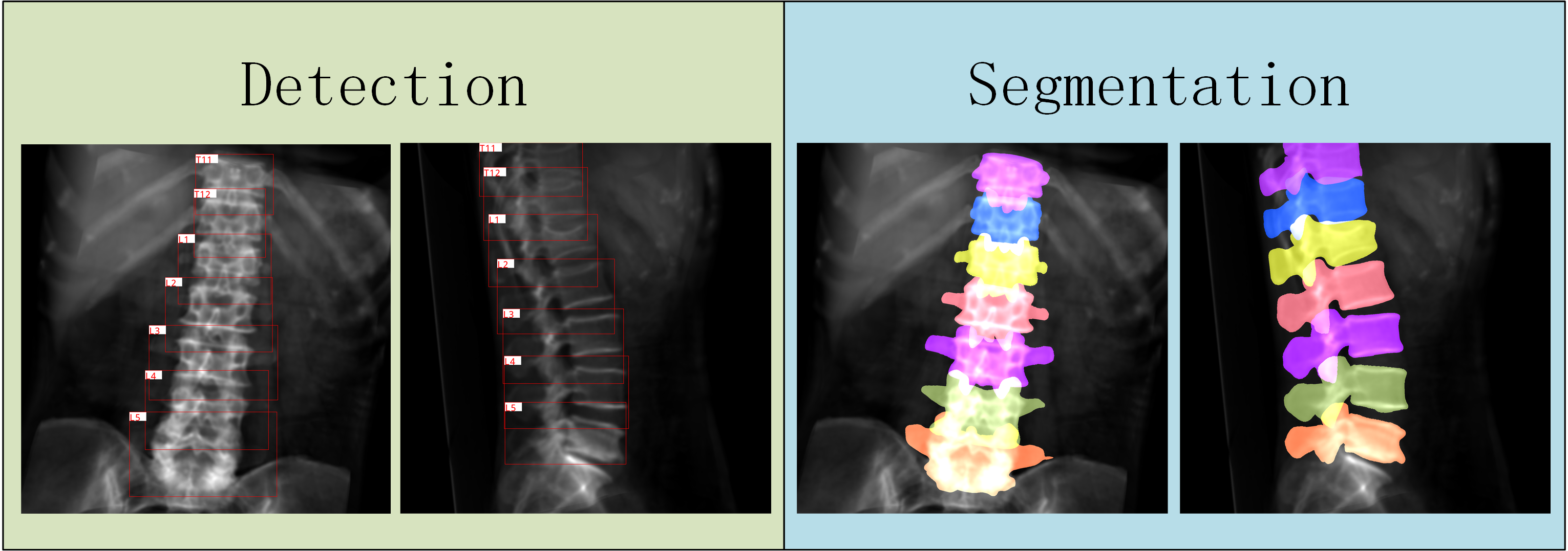
The ITK projection is used to obtain the horizontal rectangular box and the rotating rectangular box according to the boundary of the mask. At the same time, the projected mask is obtained as the segmentation label.


)T8%5B%5B4BDC1GRR%7DZ@AQ$%60CK.png)

